Cell Reaction to Surface Morphology
Roughness Feature-Size Gradients
New techniques for preparing roughness feature-size gradients are under investigation for understanding better the osteoblasts opposite reaction to different feature-size gradients (see Micro-Scale Morphological Gradients and Nano-Scale Morphological Gradients).
Polystyrene (PS) particle-height gradients were fabricated by adsorbing PS particles onto a TiO2-coated silicon wafer. The wafer was then subsequently exposed to UV light in an oxygen atmosphere. Exposing this TiO2 layer to UV light oxidises the PS particles and therefore reduce their height (see Fig. 1).

Morphological Gradients for Protein Adsorption
The success of a surgical implant is dependent on an appropriate cellular response to the implant surface [1]. Wound healing around implants is a complex process in which water molecules from the surrounding blood first get in contact with the surface. In the next step, ions and proteins will adsorb, before cells will respond to the protein-covered surface [2]. This rapid adsorption of proteins from blood acts as a translation of the implant surface into a biological language to which the cells can respond. It is crucial for later cell response [1].
Surface roughness in the micron-scale appears flat to a protein molecule, but this situation changes on the nanometre scale
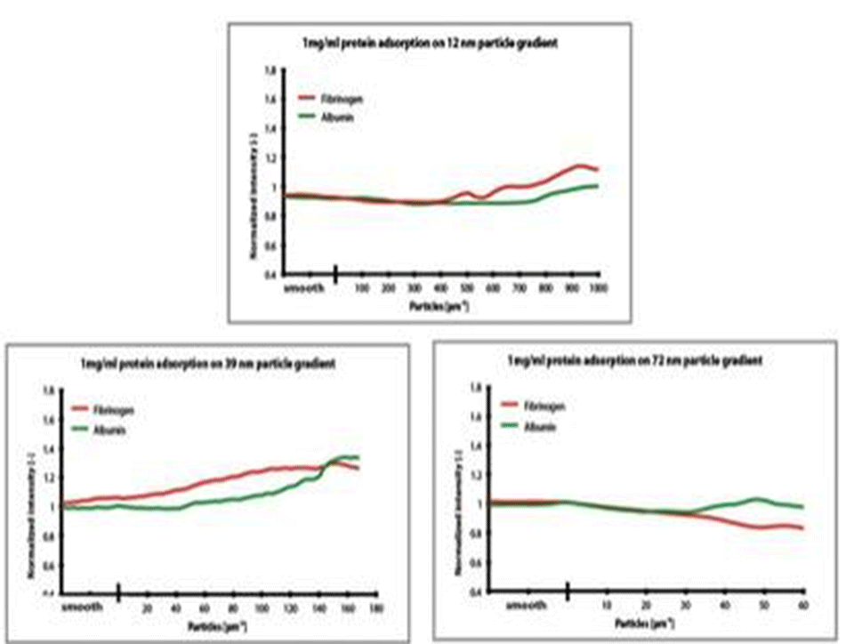
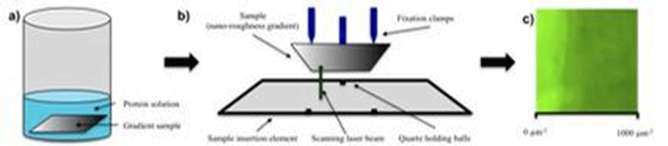
To study the effect of nano-roughness on protein adsorption, nanoparticle density gradients are fabricated (see Nano-scale Morphological Gradients). In order to mimic the surface of implants, the samples are sputter coated with titanium. Finally, the gradients are exposed to fluorescently labelled albumin and fibrinogen. To achieve an easy and rapid data read-out, a fluorescence micro-array scanner is used to map both fibrinogen and albumin (see Fig. 2).
Particles of different sizes have been tested, with diameters of 12, 39 and 72 nm. The protein-adsorption experiments have shown no significant influence of nano-features on protein adsorption for all sizes of nanoparticles investigated (see Fig. 3).